11 August 2022: Clinical Research
Surgical Management of 48 Patients with Retrosternal Goiter and Tracheal Stenosis: A Retrospective Clinical Study from a Single Surgical Center
Tao Zuo 1234ADEFG* , Zhaoming Gao 1345BC* , Zhiguo Chen 2BC* , Bin Wen 13BC , Baojun Chen 2CDF , Zhenfa Zhang 134ADEF*DOI: 10.12659/MSM.936637
Med Sci Monit 2022; 28:e936637
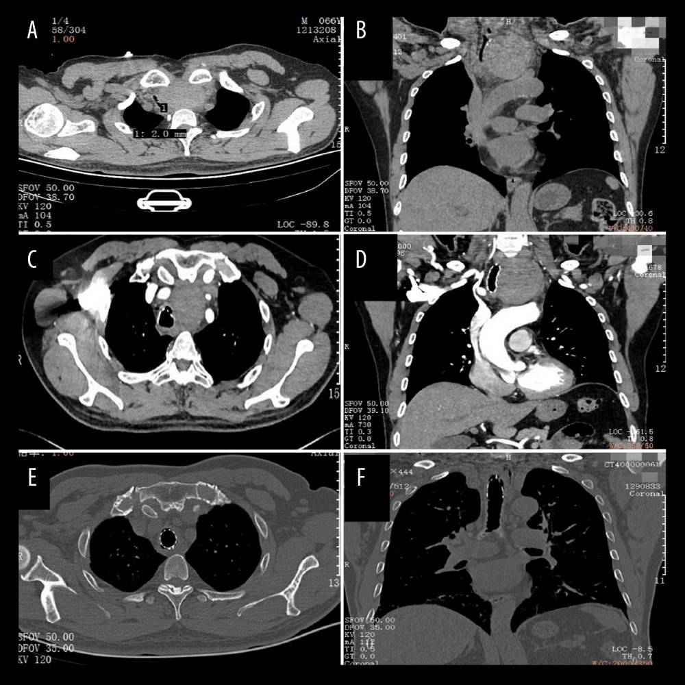
Figure 4 A patent of CT images before and after operation. The CT findings from a 66-year-old male patient revealed a type 1 RG with grade III tracheal stenosis. Axial (A) and coronal (B) images in soft-tissue windows are shown. Axial (C) and coronal (D) images in soft-tissue windows are shown after tracheoscopy. Intraoperative frozen sections suggested benign lesions. Axial (E) and coronal (F) images in soft-tissue windows are shown after surgery.