04 December 2021: Clinical Research
A Pilot Randomized Controlled Trial of Intermittent Theta Burst Stimulation as Stand-Alone Treatment for Post-Stroke Aphasia: Effects on Language and Verbal Functional Magnetic Resonance Imaging (fMRI)
Jane B. Allendorfer 1BCDEF* , Rodolphe Nenert 1BCDE , Jennifer Vannest 2ABCDE , Jerzy P. Szaflarski 13ABCDEFGDOI: 10.12659/MSM.934818
Med Sci Monit 2021; 27:e934818
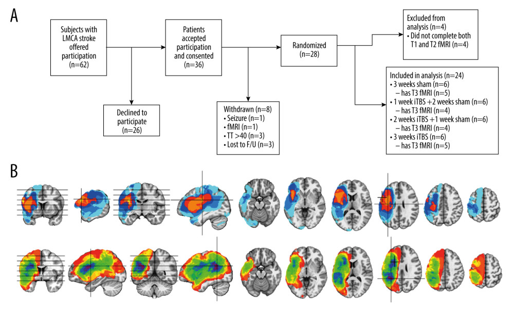
Figure 1 Overview of CONSORT flow diagram (A) and composite lesion maps for the iTBS treatment groups (B). (A) The diagram outlines recruitment of subjects who suffered a left middle cerebral artery (LMCA) stroke and how many participants did and did not complete both fMRI visits at baseline within 1 week of iTBS treatment initiation (T1), within one week of treatment completion (T2), and again after 12 weeks following treatment completion (T3). (B) The composite lesion map color scale for the sham iTBS group (top; n=6) ranges from the minimum (n=1 in light blue) to the maximum (n=6 in yellow) number of participants that show overlap of lesions in 2 locations indicated by the crosshairs in the sagittal and axial slices. The composite lesion map color scale for the active iTBS group (bottom; n=18) ranges from the minimum (n=1 in dark orange) to the maximum (n=14 in maroon) number of participants that show overlap of lesions in 2 locations indicated by the crosshairs in the sagittal and axial slices. Left in the image is left in the brain. The inferior to superior horizontal lines on the coronal images indicates each axial slice from left to right.