11 April 2022: Clinical Research
High-Resolution Magnetic Resonance Imaging (HR-MRI) Evaluation of the Distribution and Characteristics of Intra-Aneurysm Thrombosis to Improve Clinical Diagnosis of Thrombotic Intracranial Aneurysm
Yan Gu 1ABCEG* , Chongchang Miao 1ABCDF* , Aimin Li 2ABDF* , Yonggang Zhang 1BD , Jian Xu 1CDFDOI: 10.12659/MSM.935613
Med Sci Monit 2022; 28:e935613
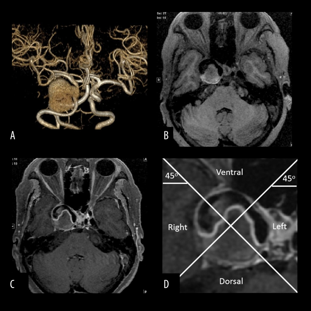
Figure 1 A thrombi aneurysm in the cavernous sinus segment of the right internal carotid artery of a 67-year-old woman. (A) Three-dimensional time of flight magnetic resonance angiography (3D-TOF-MRA) showing an aneurysm in the cavernous sinus segment of the right internal carotid artery with a size of 3.4 cm and aspect ratio value of 4.1, the shape of the aneurysm is irregular, and filling defects are seen in the aneurysm. (B) Axial T1WI image from the Philips 3D high-resolution magnetic resonance imaging (HR-MRI) scanner showing irregular isointense thrombosis in the aneurysm. (C) Axial T1WI enhanced image from the Philips 3D HR-MRI scanner showing diffused enhancement of the aneurysm wall and thrombus surface. (D) Enlarged view of the aneurysm in (C) showing the aneurysm divided into 4 quadrants (left, right, ventral, and dorsal) with 2 intersecting lines at the center of the aneurysm, and the thrombus located on the dorsal side of the aneurysm. All images were analyzed by GE adw 4.5 post-processing workstation.