08 August 2022: Clinical Research
Identification of Hepatic Dendritic Cells in Liver Biopsies in Patients with Metabolic Dysfunction-Associated Fatty Liver Diseas (MAFLD) and Obesity
Beatriz Barranco-Fragoso 12ABC , Shreya C. Pal 34CDEFG , Luis E. Díaz-Orozco 34ABCDEF , Rita Dorantes-Heredia 5BCD , Xingshun Qi 6F , Nahum Méndez-Sánchez 34ACDEF*DOI: 10.12659/MSM.937528
Med Sci Monit 2022; 28:e937528
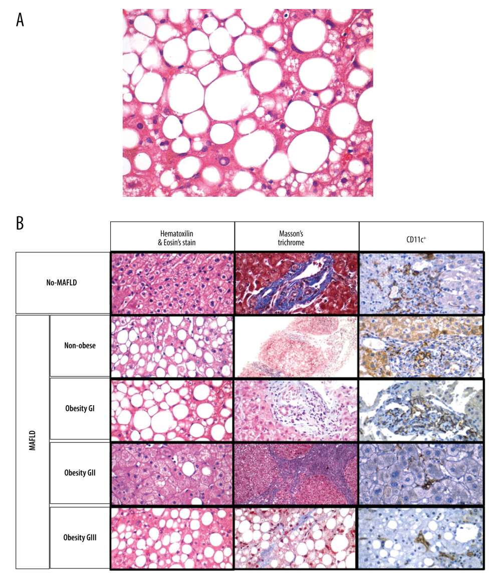
Figure 3 (A) A photomicrograph of the liver biopsy from a 54-year-old woman with metabolic dysfunction-associated fatty liver disease (MAFLD)The histology of the liver biopsy shows steatosis with round vacuoles in the liver cells where lipid has been removed during tissue processing. Note the lack of inflammation or fibrosis in this biopsy, although there may be areas of increased mononuclear cells. No necrosis is seen. Hematoxylin and eosin (H&E). Magnification ×40. (B) Comparison of liver biopsy (stained by hematoxylin and eosin, and Masson’s trichrome stain, as well as CD11c+ expression) from individuals with no metabolic dysfunction-associated fatty liver disease (MAFLD) and from patients with MAFLD in different categories according to body mass index (BMI). Based on different stains, it is possible to see the expression of dendritic cells (last row, as brown colored) varies in number and in distribution. Furthermore, the histological differences on the degree of lipid droplet saturation as well as fibrosis (with both hematoxylin and eosin, as with Masson’s stain) can be observed.