01 December 2022: Clinical Research
Clinical Outcomes of Extracranial Carotid Artery-Related Stroke Eligible for Mechanical Reperfusion on Top of Per-Guidelines Thrombolytic Therapy: Analysis from a 6-Month Consecutive Patient Sample in 2 Centers
Karolina Dzierwa 12ABDEF* , Magdalena Knapik 234ABDEF , Łukasz Tekieli 235ABDEF , Adam Mazurek 23ABDEF , Małgorzata Urbańczyk-Zawadzka 26BCD , Artur Klecha 7ABD , Tomasz Kowalczyk 7ABD , Teresa Koźmik 7ABD , Łukasz Wiewiórka 2567ABD , Paweł Banyś 6BCD , Ewa Węglarz 25BC , Justyna Stefaniak 8CD , Rafał T. Nizankowski 9ADF , Iris Q. Grunwald 1011ADEF , Piotr Musiałek 23ABCDEF*DOI: 10.12659/MSM.938549
Med Sci Monit 2022; 28:e938549
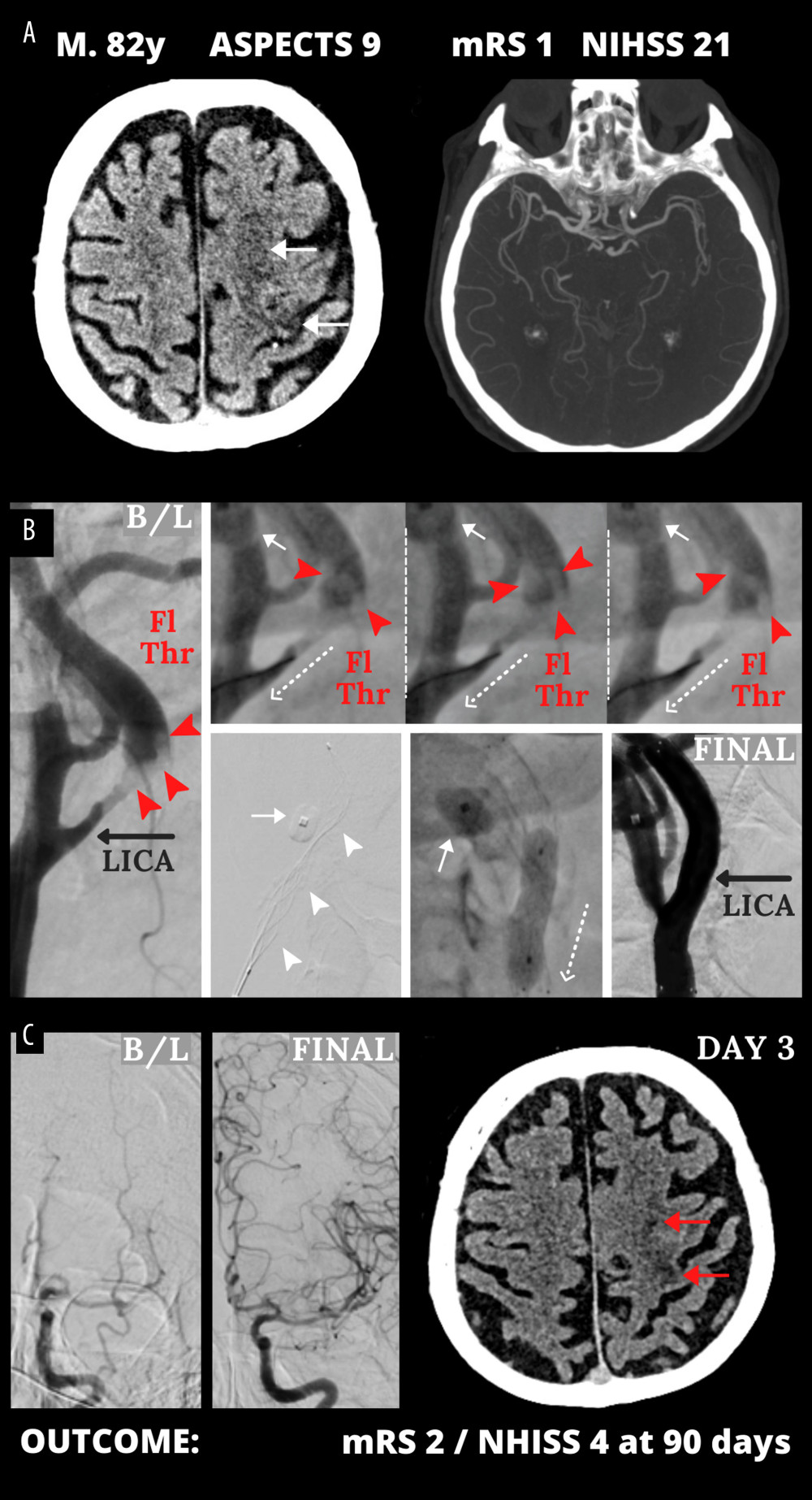
Figure 2 Emergency mechanical reperfusion in acute carotid artery origin ischemic stroke. (A), (left panel) – mild vascular changes (white arrows) seen on admission cerebral computed tomography Alberta Stroke Program Early Computed Tomography Score (ASPECTS) 9 in a patient who was emergency-transferred to a cardioangiology-based Thrombectomy-Capable Stroke Center from external Neurology; (right panel) – diminished flow to the left hemisphere on computed tomography angiography (compare left vs right). (B) Catheter angiography – left internal carotid artery (LICA) near-occlusion with a floating thrombus (Fl Thr, red arrowheads) and next stages of emergency mechanical reperfusion (from left to right, upper and lower panels): transient flow reversal (dotted arrows; enhanced by active aspirations at the procedure critical steps) – proximal cerebral protection device – Mo.Ma (Medtronic, Tolochenaz, Switzerland), external carotid artery balloon (white arrow). Following thrombectomy (carotid-dedicated adjustable-diameter stentriever – TigerTrieverXL (Rapid Medical, Yokneam, Israel), the culprit lesion was sequestrated (white arrowheads), using a micronet-covered stent – C-Guard (InspireMD, Tel Aviv, Israel) with post-dilatation embedding. (C), (left panel) – effective lumen reconstruction resulted in normalized left hemispheric cerebral blood supply symptoms regressed, (right panel) – discharge cerebral computed tomography showed a minor cerebral infarct (red arrows). B/L indicates baseline, (mag) = magnified image. Figure was created with the use of Canva (Perth, Australia).